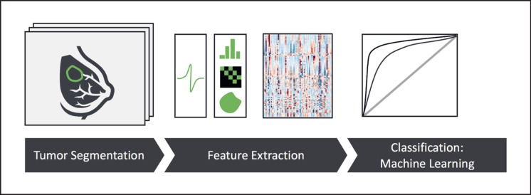
Fig. 1.
Outline of a state-of-the-art radiomics analysis [88]. Briefly, a tumor is outlined or contoured on tomographic images. In the next step, a range of quantitative image features is computed, typically comprising shape features, signal intensity distributions, and image texture. Finally, machine learning is used to establish a link between this high-dimensional data space and the relevant clinical endpoint or outcome.

