. 2019 Jun 25;12(2):233–235. doi: 10.3400/avd.cr.18-00158

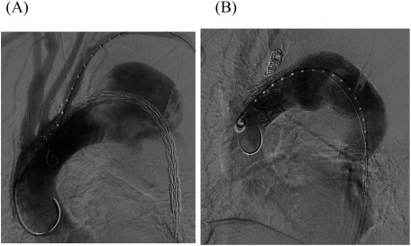
Fig. 2 (A) Intraoperative angiography showing a thoracic aortic aneurysm close to the left subclavian artery (LSA). The need for LSA coverage is apparent. (B) The proximal stent was 45 mm in diameter and 200 mm long; the distal stent was 45 mm in diameter and 150 mm long. LSA coverage was required to achieve an adequate proximal seal. Additionally, LSA coil occlusion was performed.

Fig. 2 (A) Intraoperative angiography showing a thoracic aortic aneurysm close to the left subclavian artery (LSA). The need for LSA coverage is apparent. (B) The proximal stent was 45 mm in diameter and 200 mm long; the distal stent was 45 mm in diameter and 150 mm long. LSA coverage was required to achieve an adequate proximal seal. Additionally, LSA coil occlusion was performed.