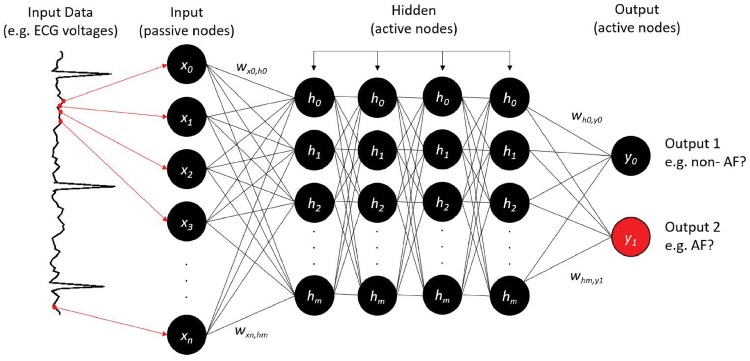
Figure 3.
Neural network design to classify atrial fibrillation from the electrocardiogram. Continuous electrocardiogram voltage points (red dots, arrows) are fed to ‘input neurons’ (x0, x1, x2, …, xm), which are coded as software objects. Hidden neurons within this three-layer network (h0, h1, h2, …, hn) connect input and output layer neurons (here, two neurons) by numerical weights (w). Deep learning typically uses multiple hidden layers, as shown here. The output indicates atrial fibrillation (y1; correct, red) or non-atrial fibrillation (y0). If the output is correct for that electrocardiogram input, weights are strengthened; else they are reduced. This process is iterated during training on multiple input electrocardiograms. The trained network can then be tested on new (unseen) electrocardiograms. Other designs could accept categorical variables (age, gender) or mixed data types.

