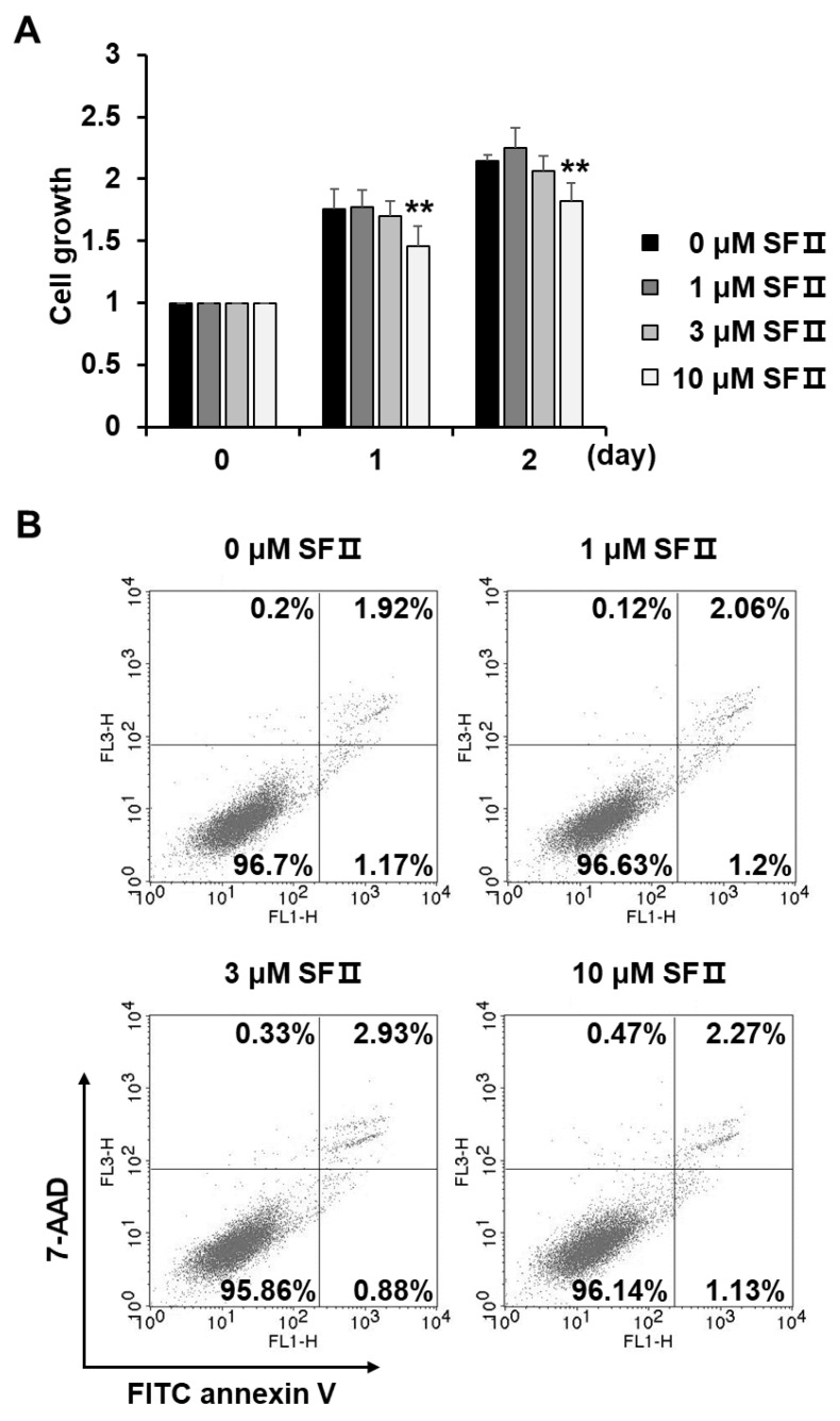
Figure 2.
Effect of skullcapflavone II on proliferation and cytotoxicity of foreskin fibroblasts. Sub-confluent foreskin fibroblasts were plated in DMEM containing 10% FBS and incubated with indicated concentrations of skullcapflavone II. (A) The number of viable cells was measured based on the absorbance at a wavelength of 565 nm using 3-(4,5-dimethyl thiazol-2-yl)-2,5-diphenyltetrazolium bromide (MTT) reagent. The number of viable cells in the presence of skullcapflavone II is shown as fold change relative to that in the absence of skullcapflavone II. ** p < 0.01 vs. the sample incubated with 0 μM skullcapflavone II. (B) After 24 h of incubation with skullcapflavone II, apoptotic cells were stained with fluorescein isothiocyanate (FITC) annexin V and 7-aminoactinomycin (7-AAD) and then analyzed by flow cytometry.

