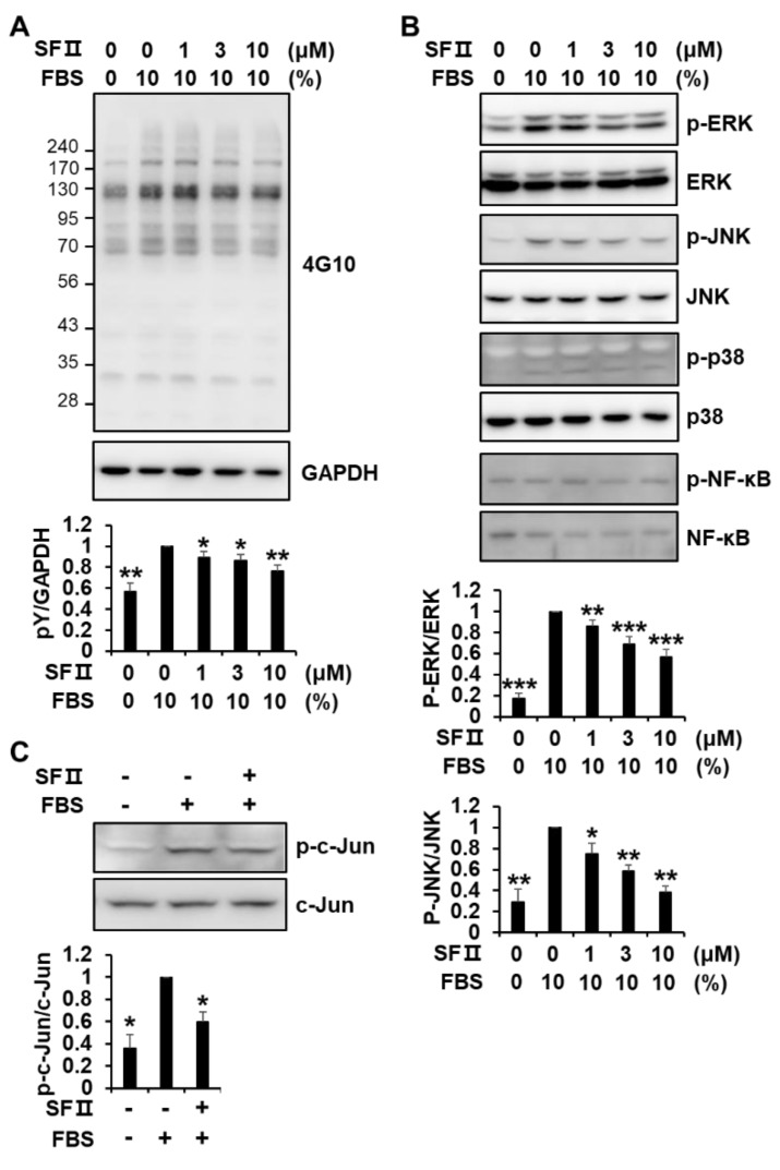
Figure 4.
Effect of skullcapflavone II on phosphorylation of signaling molecules in foreskin fibroblasts. Sub-confluent foreskin fibroblasts were starved for 24 h. (A,B) Cells were pre-incubated for 30 min with indicated concentrations of skullcapflavone II and then stimulated for 10 min with 10% FBS. Cell lysates were subjected to 9% SDS-PAGE and Western blot analysis with anti-phosphotyrosine (4G10) (A), anti-phospho-extracellular signal-regulated kinase (ERK), anti-phospho-c-Jun N-terminal kinase (JNK), anti-phospho-p38 mitogen-activated protein kinases (MAPK), and anti-phospho-nuclear factor kappa light chain enhancer of activated B cells (NF-κB) p65 antibodies (B). (C) Cells were pre-incubated for 30 min with (+) or without (−) 3 μM skullcapflavone II, and then FBS was added to a final concentration of 10% and incubated for 30 min. Cell lysates were analyzed by Western blotting with anti-phospho-c-Jun and anti-c-Jun antibodies. * p < 0.05, ** p < 0.01, and ***p < 0.001 vs. the sample incubated with 10% FBS alone.

