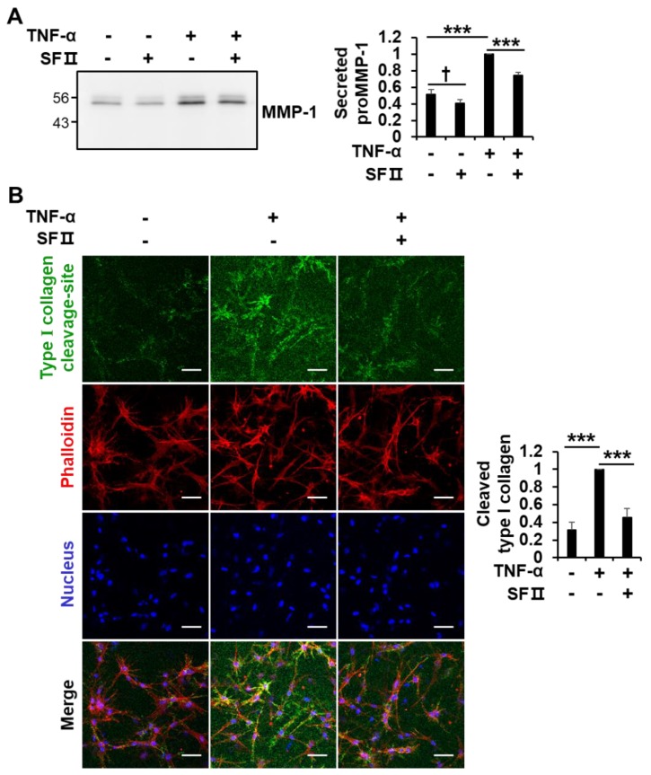
Figure 6.
Effect of skullcapflavone II on TNF-α-induced type I collagen degradation in 3D culture of foreskin fibroblasts. Foreskin fibroblasts were embedded within a 3D type I collagen matrix. After polymerization for 1 h at 37 °C, cells embedded in the collagen matrix were incubated for 24 h in serum-free DMEM with (+) or without (-) 3 μM skullcapflavone II and with (+) or without (−) 1 ng/mL TNF-α. (A) The conditioned medium in the 3D culture was analyzed by 9% SDS-PAGE and Western blot with anti-MMP-1 antibody. *** p < 0.001 vs. the sample incubated with TNF-α alone; † p < 0.05 vs. the sample incubated without TNF-α and without skullcapflavone II (B) The 3D matrix containing foreskin fibroblasts was stained with anti-type I collagen cleavage-site antibody and Alexa Fluor® 488 goat anti-rabbit IgG (H+L), phalloidin–rhodamine and Hoechst 33258, and cells were analyzed by confocal fluorescence microscopy (200×). Scale bar, 50 μm. The quantification of cleaved type I collagen (Alexa Fluor® 488) relative to nuclear staining (Hoechst 33258) obtained from six randomly chosen fields is shown as the mean ± S.D. *** p < 0.001 vs. the sample incubated with TNF-α alone.

