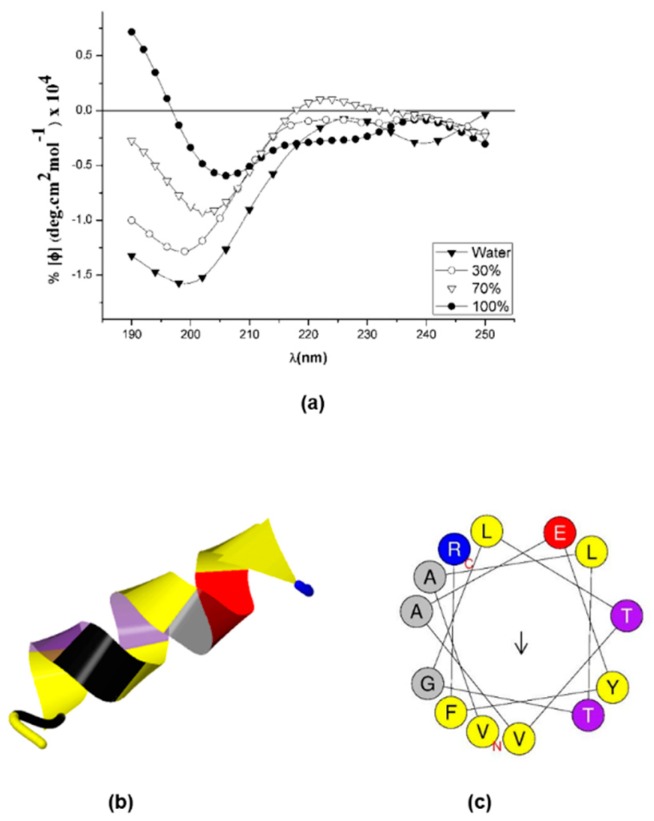
Figure 5.
Sarconesin II secondary structure. (a) A Jasco-1500 instrument was used for measuring sarconesin II’s circular dichroism spectrum. Sarconesin II CD spectra variation at 0%, 30%, 70% and 100% trifluoroethanol (TFE) concentrations (b). I-TASSER sarconesin II secondary structure gave an α-helix, depicted in spiral ribbon format, using common colors. (c) Computation of sarconesin II α-helical wheel [54]. No hydrophobic face reported. Note slightly opposite arrangement of hydrophobic (yellow) versus charged (red, blue) aa.

