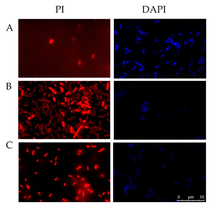
Figure 8.
Fluorescence microscopy of E. coli cells incubated for 4 h at 37 °C and stained with PI [56] or DAPI (blue). Untreated control cells, PBS (A), cells treated with sarconesin II (B), cells treated with ampicillin for PI or ciprofloxacin for DAPI (C). PI assay revealed bacterial membrane alteration when treated with sarconesin II and DAPI-stained cells had partial fluorescence, showing DNA fragmentation by sarconesin II.

