FIG 1.
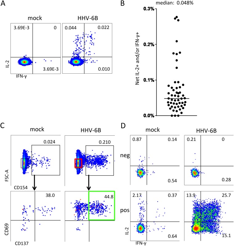
Isolation and enrichment of HHV-6B-specific CD4 T cells. (A) PBMCs from a representative donor were stimulated for 18 h with mock or HHV-6B lysate and tested by intracellular cytokine staining (ICS) for IL-2 and IFN-γ expression. Gated CD3+ and CD4+ cells are shown. (B) Results from similarly tested ex vivo PBMCs of 53 donors. For each, HHV-6B-treated cells expressing either cytokine, or both, were totaled, and mock values were subtracted for the net IL-2 and/or IFN-γ HHV-6B-specific T-cell frequency. The horizontal bar is the median (0.048%). (C) Sorting scheme for HHV-6B-specific T cells. Live cells from the same donor as in panel A were gated for CD3 and CD4 expression; from these, cells expressing CD154, CD69, and CD137 (green) were sorted and expanded in culture for downstream assays. Cells negative for CD154 (red gate) were sorted as a negative control. FSC, forward scatter. (D) Expanded polyclonal T cells from the same donor as in panel A originating from CD154-negative cells (“neg”) or CD154/CD137/CD69 triple-positive cells (“pos”) were evaluated to document enrichment for HHV-6B specificity among the AIM-positive cells. Numbers are percentages of gated cells expressing the indicated pattern of cytokines or activation markers.

