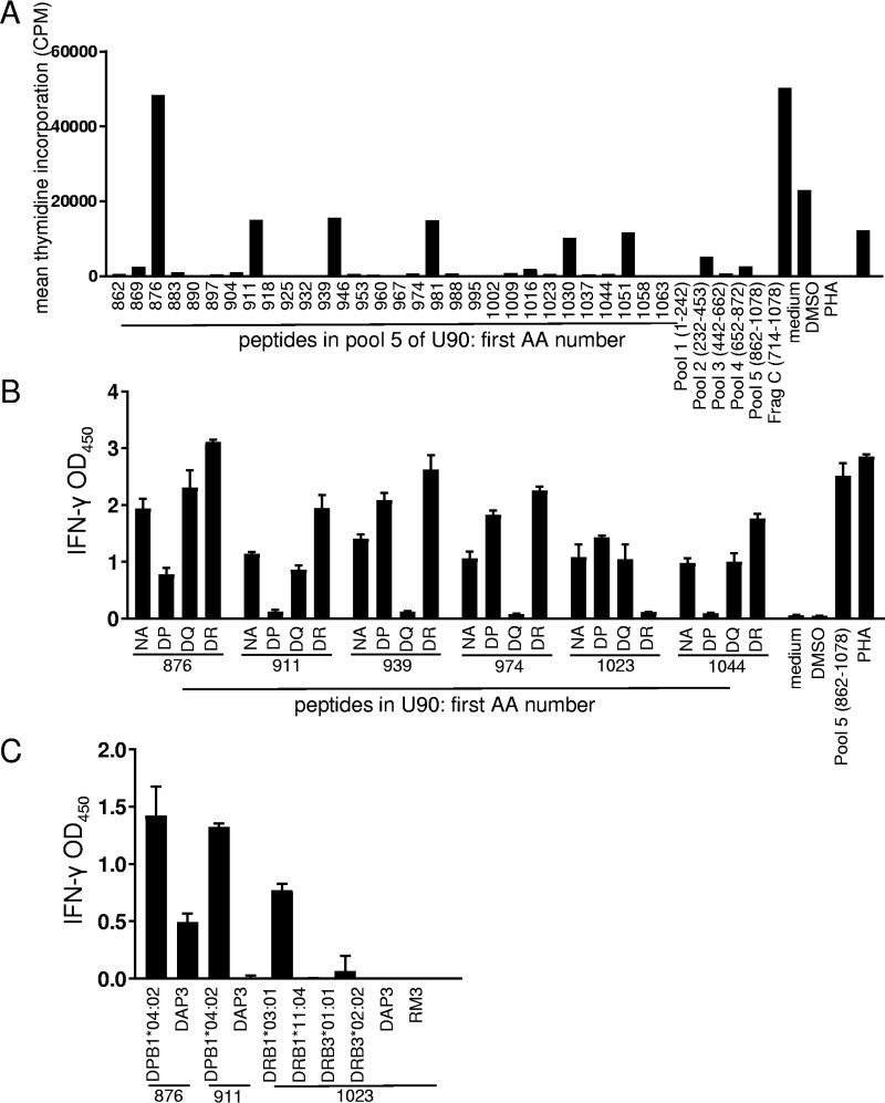
FIG 3.
Detection of CD4 T-cell epitopes in HHV-6B U90. (A) Polyclonal HHV-6B-reactive CD4 T cells from a representative donor were tested against U90 peptides. Controls at the right include responses to U90 peptide pools and the U90 fragment C IVTT polypeptide, which corresponds to part of pool 4, and pool 5. Numbers on the x axis indicate starting amino acid (AA) positions of 18-mer peptides or amino acid positions covered by peptide pools. Values are means of data from duplicates. DMSO, dimethyl sulfoxide. (B) Reactive U90 peptides were retested with HLA locus-specific blocking monoclonal antibody specific for HLA-DP, HLA-DQ, or HLA-DR or no antibody (NA). After overnight stimulation, supernatants were tested for IFN-γ by an ELISA. Values are means and standard deviations of data from triplicates. OD450, optical density at 450 nm. (C) Selected peptides from panel B were retested using single-HLA class II-expressing cell lines as APCs with ELISA readouts in triplicate. U90 peptides are indicated at the bottom. HLA class II-negative parental cell lines DAP3 and RM3 were used as controls.

