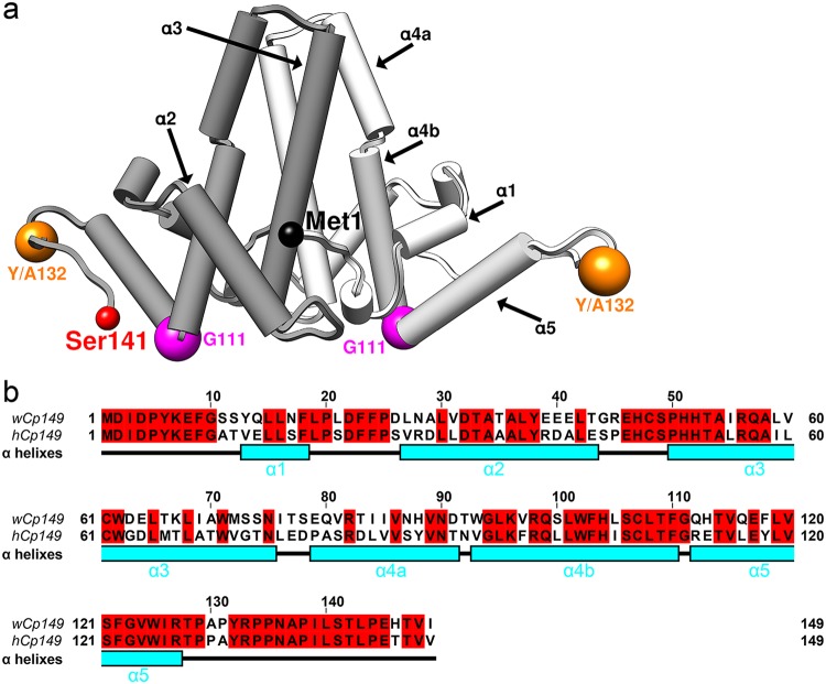
FIG 1.
WHV and HBV have similar assembly domains. (a) A capsid protein dimer is represented using pipes and planks with the two monomers shown in different shades of gray. The positions of key residues discussed in text, Y132 and G111, are marked by spheres. The first residue, Met1, of one monomer and the end residue solved in crystal structure, Ser141, of the other monomer are also indicated. (b) Sequence alignment shows that wCp149 and hCp149 have 63% sequence identity. The sequence is correlated with α-helixes (cyan), numbered as in panel a.

