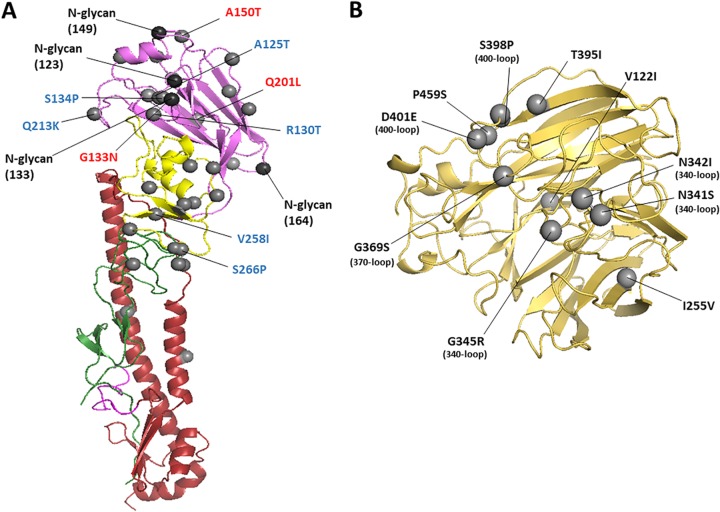
FIG 5.
The hemagglutinin (HA) and neuraminidase (NA) structure of A/CK/PB/16 virus. The amino acid changes from A/CK/JL/12 to A/CK/PB/16 are shown as gray spheres using H7 numbering and N3 numbering. (A) Ribbon diagram of H7 HA. Only a monomer is shown. Potential vaccine escape mutations and residues related to chicken and duck adaptation are labeled in red and blue, respectively. The HA1 is depicted in green (F′ subdomain), yellow (vestigial esterase subdomain), and violet (receptor binding domain). The HA2 is shown in magenta (fusion peptide) and red (F subdomain). The amino acid changes from A/CK/JL/12 to A/CK/PB/16 are represented as gray spheres. The possible glycosylation sites are represented as black spheres. (B) Ribbon diagram of the head region of N3 NA. Only one monomer is shown. Amino acid changes located in the 340-, 370-, and 400-loop regions are marked in parentheses.

