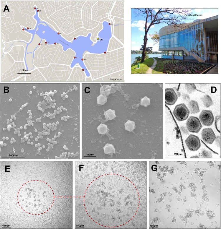
FIG 1.
Faustovirus mariensis isolation sites, particle images, and cytopathic effects. (A) Pampulha Lagoon map with collection sites highlighted (dots). The yellow dot represents where F. mariensis was collected, in front of the Pampulha Art Museum (top right of photo). Map courtesy of Google Maps. (B to D) F. mariensis viral particles visualized by scanning (B and C) and transmission (D) electron microscopy. (E to G) Plaque-forming unit (PFU) induced by F. mariensis infection in a Vermamoeba vermiformis monolayer. (F) Closeup of a PFU shown in panel E, observed 24 h postinfection. (G) Forty-eight hours postinfection, the PFUs expand and coalesce.

