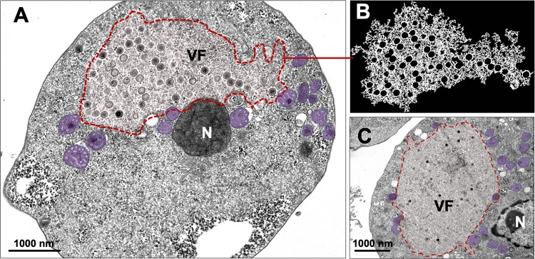
FIG 3.
Electron-lucent viral factory and cytoplasmic modifications induced by Faustovirus mariensis. (A to C) F. mariensis presents an electron-lucent viral factory (contoured in red and shown in detail in panel B), which was not easily distinguished from the rest of the cytoplasm and was observed at the perinuclear region. It is possible to visualize the abundant presence of mitochondria surrounding the viral factory (purple highlights in panels A and C). VF, viral factory; N, nucleus. Image B was obtained by TEM and graphically highlighted by using IOS image visualization software (Apple Technology Company).

