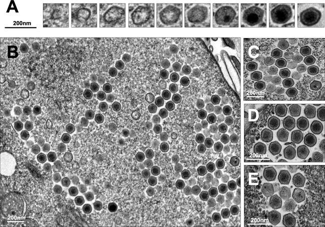
FIG 4.
Faustovirus mariensis morphogenesis and particle organization in honeycomb-like structures. (A) F. mariensis morphogenesis begins with crescents, open structures of approximately 50 nm, which grow as an electron-dense material of the viral factory fills them. Particles of almost 200 nm without genomic content are observed in late phases of morphogenesis, when the genome is incorporated and centralized within several newly formed viral particles. (B to E) F. mariensis progeny are organized in a honeycomb fashion inside viral factories. Small honeycombs expand as new mature viruses are formed and coalesce to others in the cytoplasm (B).

