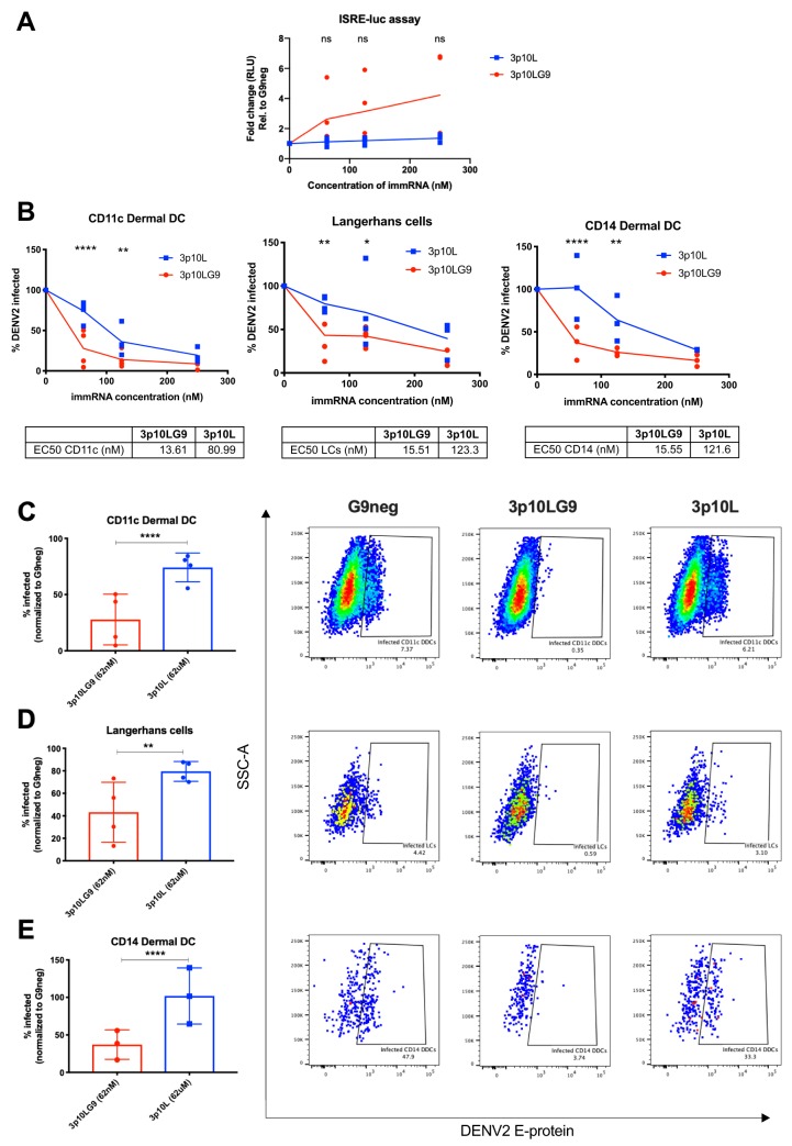
FIG 6.
3p10LG9 has a higher efficacy than 3p10L as a prophylaxis against DENV-2 infection in primary human skin DCs. (A) Human skin DCs were transfected with 250 nM, 125 nM, and 62 nM 3p10LG9 or 3p10L and incubated for 24 h. Supernatant was incubated on HEK-293T cells containing a luciferase reporter driven by an interferon-stimulated response element (ISRE-luc), and luminescence was measured after 6 h. Results are presented as fold change compared to G9neg. Each symbol represents a sample from one donor. Statistical significance (P < 0.05) was determined using a two-way ANOVA with Dunnett’s multiple-comparison test (ns, not significant). (B) Human skin DCs transfected with 250 nM, 125 nM, and 62 nM 3p10LG9 or 3p10L for 24 h were infected with DENV-2 at an MOI of 5 for 48 h. Flow cytometry was used to quantify the percentage of DENV-2-infected cells in each subpopulation of skin DCs by intracellular staining with 4G2 antibody. The percentage of cells infected for each condition was normalized to G9neg. (C to E) Percentage of infected cells for each subpopulation of skin DCs taken from the data plotted in panel B after transfection with 62 nM 3p10LG9 or 3p10L (left), and representative flow cytometry (right). (C) CD11c DDCs; (D) Langerhans cells; (E) CD14 DDCs. Each symbol represents one donor. Bars show means ± SD. Statistical significance (P < 0.05) was determined using a two-way ANOVA with Dunnett’s multiple-comparison test (*, P ≤ 0.05; **, P ≤ 0.01; ****, P ≤ 0.0001).

