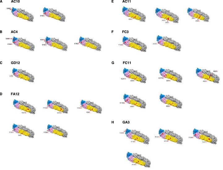
FIG 4.
Escape mutants are mapped to a crystal structure of the ZIKV E protein. Panels A to H show graphical representations of the envelope protein with relevant mutations found on plaque-purified viruses 1 to 6 used for the experiment shown in Fig. 3 indicated in red. PDB accession number 5JHM was used to generate the three-dimensional model using UCSF Chimera.

