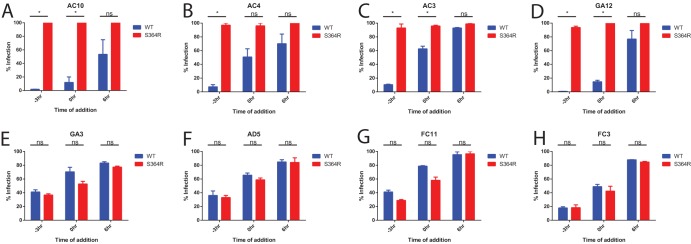
FIG 7.
Neutralization occurs at a binding step. Analysis of the timing of antibody neutralization activity was performed. The results for synchronized infections of Vero cells with wild-type or S368R MR766 virus are shown. Vero cells were equilibrated to 4°C at −3 h, and virus plus antibody or virus alone was added to the cells. At 0 h Vero cells were washed twice with PBS, warm medium was added with the relevant antibody, and cells were moved to 37°C. For each assay, results are normalized to those of infections performed without any antibody added. Antibody was added at a concentration of 10 IC50s as determined by plaque reduction neutralization test. Infection was measured by 4G2 anti-envelope staining at 48 h postinfection. Assays were performed in duplicate, and error bars represent SEM. *, P < 0.05; ns, not significant.

