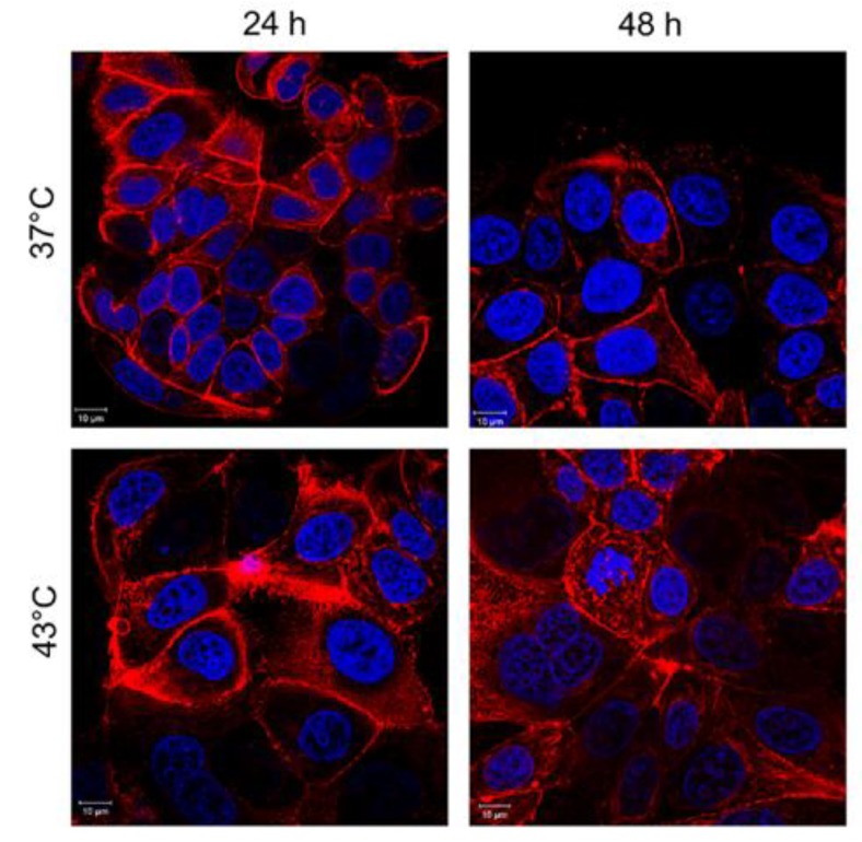
Figure 8.
Effects of temperature on actin network of MCF-7 cells after 24 h and 48 h. Cells were fixed and then stained with Phalloidin–ATTO 488 and DAPI. The 2D images of cortical actin were acquired by a Zeiss LSM700 (Zeiss) confocal microscope equipped with an Axio Observer Z1 (Zeiss) inverted microscope using a ×100, 1.46 numerical aperture oil immersion lens. All data were processed by ZEN software (Zeiss).

