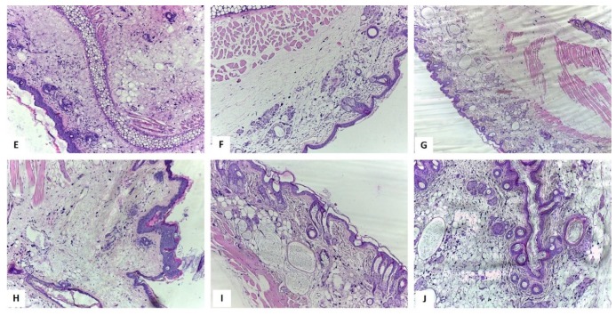
Figure 8.
Histological aspects of the skin H-E stain, E: Blank hydrogel group, ob. 10× – the sample displayed inflammatory cells distributed in the entire dermis and massive interstitial edema; F: ZnCl2 1%, ob. 10× – Reduced number of neutrophils and moderate interstitial edema; G: ZnCl2 5%, ob. 4× – Reduced number of neutrophils and dermis hyalinization; discreet edema and mild congestion of the blood vessels; H: EM2 1% group, ob. 10× – in the entire dermis mild inflammation and moderate interstitial edema were observed; I: EM2 + ZnCl2 1%, ob. 10× – Hyalinization of the dermis and epidermal alterations were similar to the group treated with ZnCl2 1%. J: EM2 + ZnCl2 5%, ob. 10× - Hyalinization of the dermis and epidermal alterations were similar to the group treated with ZnCl2 5%. [29].

