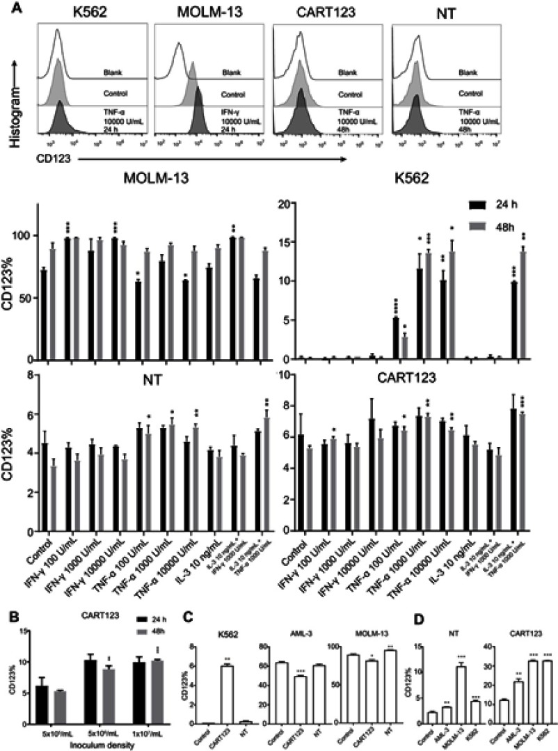
Figure 5.
Induced expression of CD123 in CAR T and myeloid leukemia cells in vitro. (A) Expression of CD123 on MOLM-13, K562, NT, and CART123 after treatment with cytokines (TNF-α, IFN-γ, and IL-3) for 24 and 48 hrs was detected by flow cytometry (mean±SEM, n=3). Data show one representative experiment. Representative plots are shown. (B) Expression of CD123 on CART123 in different density of seeding cells after in vitro culture for 24 and 48 hrs (mean±SEM, n=3). Data show one representative experiment. (C-D) CFSE-labeled effector cells (CART123 or NT) and target cells (MOLM-13, K562, and AML-3) were seeded in the upper chamber, and healthy donor-derived PBMC were seeded in the lower chamber of the in vitro co-culture model. After co-culturing for 24 hrs, target cells (C) and CFSE-labeled effector cells (D) and were analyzed for CD123 expression (mean±SEM, n=3). Data show one representative experiment. *P<0.05; **P<0.01; ***P<0.001; ****P<0.0001.
Abbreviations: IFN, interferon; TNF, tumor necrosis factor, HUVECs, human umbilical vein endothelial cells; CAR, chimeric antigen receptor; NT, non-transduced T; 7-AAD, 7-Aminoactinomycin D; AML, acute myeloid leukemia; E:T, the effector to target cell ratio; PBMC, peripheral blood mononuclear cell; CFSE, carboxyfluorescein succinimidyl ester.

