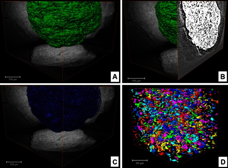
Figure 3.
SR-µCT of CHA microsphere: (A) VR of microsphere with normalized orthoprojections; (B) orthoslice showing porous space inside the sphere; (C) VR of porous space inside sphere; (D) box representing individual pores of central microsphere region.
Abbreviations: orthoslice, orthogonal slice; SR-µCT, synchrotron radiation-based X ray microtomography; CHA, carbonated hydroxyapatite; VR, volume rendering.

