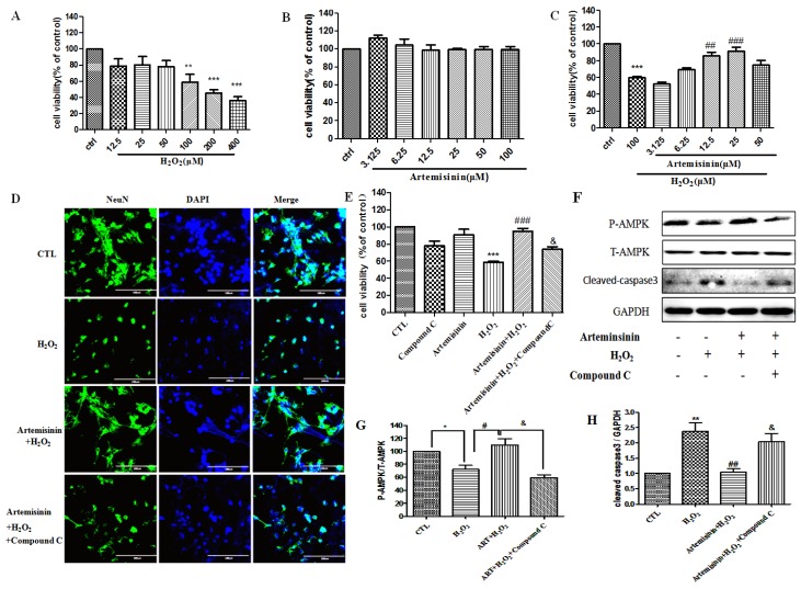
Figure 6.
Artemisinin attenuated the decrease in cell viability caused by H2O2 in neuronal cells. (A) The cytotoxicity of H2O2 on neuronal cells. (B) Dose of artemisinin. (C) Primary cultured hippocampal neurons were pretreated with artemisinin at indicated concentrations and then induced with or without 100 μM H2O2 for another 24 h, and cell viability was measured using the MTT assay. (D) Primary cultured hippocampal neurons pretreated with 2.5 μM compound C for 30 min were treated with 25 μM artemisinin for 2 h, and then incubated with or without 100 μM H2O2 for another 24 h. Immunocytochemistry of NeuN in each group was detected. The pictures were taken at a magnification of 40× (100 μm). (E) Primary cultured hippocampal neurons pretreated with 2.5 μM compound C for 30 min were treated with 25 μM artemisinin for 2 h, and then incubated with or without 100 μM H2O2 for another 24 h. Cell viability was measured by the MTT assay. (F) The expression of phosphorylated AMPK, total AMPK, cleaved caspase-3 and GAPDH were detected by western blot. (G,H) Quantitative analysis of (F). The data were represented as the mean ± SD. ** p < 0.01, *** p < 0.001, ## p < 0.01, ### p < 0.001.

