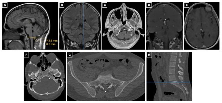
Figure 2.
Patient 2 Intracranial and Spinal Imaging. Panels (A–E): MRI of the brain. Panel (A): Sagittal post-contrast T1-weighted sequence shows the left cerebellar tonsil at the lowest tonsillar position, 8 mm through the foramen magnum. Narrow, underdeveloped clivus and occipital bone as well as uncalcified synchondroses of the odontoid are also seen (arrowheads). Panel (B): Coronal T1-weighted sequence demonstrating the location of the measurement in A. Panel (C): Axial post-contrast T1-weighted sequence showing crowding of the brainstem by the cerebellar tonsils in the foramen magnum (arrows). Panel (D): Coronal T1-weighted venous phase post-contrast sequence shows a developmental venous anomaly of the Vein of Galen confluens draining the choroid plexus through the velum interpositum (arrowheads). Panel (E): Axial T1-weighted venous phase post-contrast sequence showing the anomalous Vein of Galen confluens. Panel (F): Axial CT of the head showing petroclival dysraphism or uncalcified petroclival synchondrosis (arrowheads). Panel (G): Axial CT of lumbo-sacral spine shows occult dysraphism of S1 (arrowhead) and sacral segmentation anomalies (arrows). Panel (H): Sagittal lumbar CT demonstrating the level of the image shown in G.

