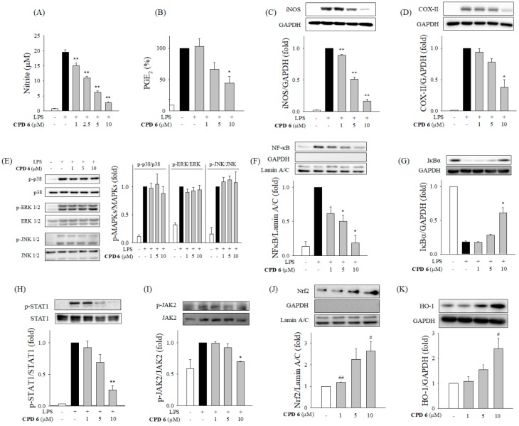
Figure 2.
Anti-inflammatory effect of CPD 6 in LPS-activated macrophages and the responsible signaling pathways. (A and B) Effects of CPD 6 on the release of NO (A) and PGE2 (B) were examined after 24 h LPS challenge in RAW 264.7 cells using the Griess reagent and ELISA, respectively. (C,D) The protein levels of iNOS (C) and COX-II (D) were measured by western blot. (E) Macrophages were stimulated by LPS for 1 h to determine phosphorylation of the MAPKs (p38, ERK, and JNK) in the presence or absence of CPD 6. (F,G) Effect of CPD 6 on NFκB nuclear translocation (F) or IκBα degradation (G) was measured after LPS treatment for 1 h 30 min, respectively. (H,I) Macrophages were challenged with LPS in the presence or absence of CPD 6 for 6 h to measure phosphorylated level of STAT1 (H) and for 2 h to measure phosphorylated JAK2 (I). (J,K) Nrf2 nuclear translocation (J) and HO-1 expression (K) were measured after CPD 6 treatment without LPS stimulation for 6 h and 24 h, respectively. GAPDH and Lamin A/C were used as internal controls for the cytoplasmic and nuclear fractions, respectively. Data are expressed as mean ± SEM, * p < 0.05, ** p < 0.01 vs. LPS-treated group. # p < 0.05, ## p < 0.01 vs. control group. N = 3 or 4.

