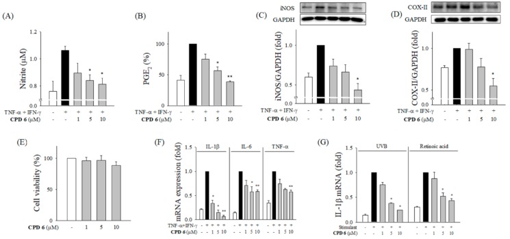
Figure 3.
Attenuation of inflammatory signals in CPD 6 in keratinocytes challenged by TNF-α and IFN-γ, UV irradiation, or a chemical irritant. (A and B) Effect of CPD 6 on the release of NO (A) and PGE2 (B) was measured after 24 h challenge of TNF-α+IFN-γ combination (10 ng/mL each) in human keratinocytes, HaCaT cells. The released amount of NO or PGE2 was determined by Griess reaction or ELISA, respectively. (C,D) The protein levels of iNOS (C) or COX-II (D) were measured in HaCaT cells activated by TNF-α/IFN-γ. (E) Effects of CPD 6 on the cell viability of HaCaT cells were examined by MTT assay. (F) The levels of mRNA for IL-1β, IL-6, and TNF-α were measured in human keratinocytes stimulated by TNF-α/IFN-γ using quantitative real time PCR. (G) The mRNA levels of IL-1β in keratinocytes were examined at 6 h after UVB irradiation (25 mJ/cm2) or exposure to retinoic acid (50 µM). (G) Data are expressed as mean ± SEM, * p < 0.05, ** p < 0.01 vs. TNF-α/IFN-γ or UVB or retinoic acid-treated group. N = 3.

