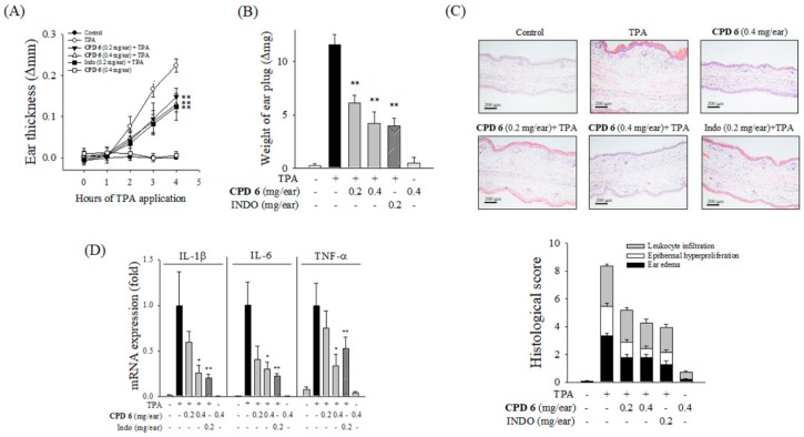
Figure 4.
Attenuation of TPA-induced acute skin inflammation by CPD 6 in mice. CPD 6 (0.2 mg or 0.4 mg) or indomethacin (INDO, 0.2 mg) was topically administered to the right ear at 30 min prior to TPA application. (A,B) The induction of ear edema was assessed by measuring ear thickness (A) and ear disc weight (B). (C) The right ear tissues were examined with H&E staining. Representative histological images from each group were shown (upper panel) and histological scores were plotted (lower panel). Black, white, and gray bars represent ear edema, epithermal hyperproliferation, and leukocyte infiltration, respectively. (D) The mRNA levels of IL-1β, IL-6, and TNF-α in the right ear tissue were determined 4 h after TPA application. Data are expressed as mean ± SEM, * p < 0.05, ** p < 0.01 vs. TPA-treated group. N = 5.

