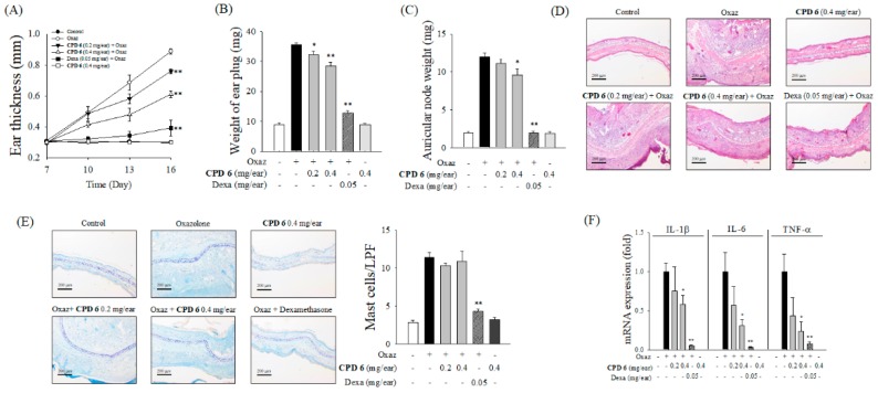
Figure 5.
Anti-inflammatory effect of CPD 6 in oxazolone (Oxaz)-induced chronic dermatitis mouse model. On day 0, sensitization was initiated by topical oxazolone treatment on dorsal skin of BALB/c mice, and then inflammatory reaction was induced by challenge with topical oxazolone application on the right ear on days 7, 10, 13, and 16. Thirty minute before and 3 h after each elicitation, CPD 6 (0.2 or 0.4 mg) or dexamethasone (Dexa, 0.05 mg) was applied to the right ear. (A to C) The changes in ear thickness (A), the weight of ear plug (B), or auricular lymph node (C) were examined at 6 h after the final oxazolone challenge. (D) Representative images of H&E staining of right ear tissues from each group are shown (upper panel) with histological scores (lower panel). Scores of ear edema, epithermal hyperproliferation, and leukocyte infiltration were expressed as black, white, and gray bars, respectively. (E) Representative histological images of right ear tissue in TB-staining were shown (left panel) and the mast cell counts were plotted (right panel). (F) The mRNA levels of pro-inflammatory cytokines in right ear tissues were determined 6 h after the final oxazolone application. Data are expressed as mean ± SEM, * p <0.05, ** p < 0.01 vs. oxazolone-treated group. N = 5.

