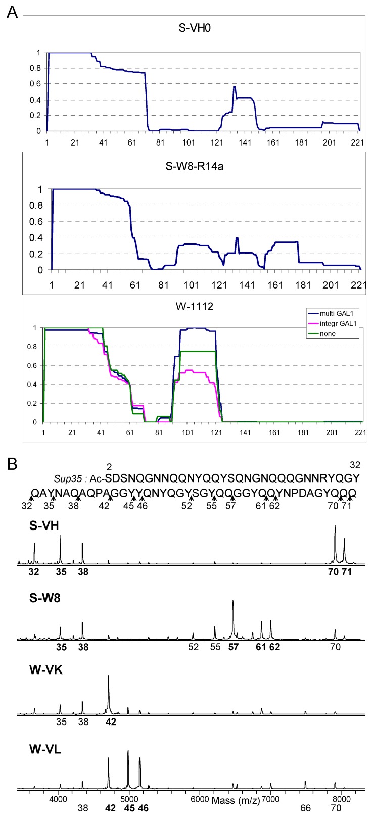
Figure 2.
Structural analysis of selected [PSI+] variants. (A) PK resistance index for the Sup35 region 2–222 of variants L20, S-W8-R4, and W-1112, calculated as described in the text. In contrast to Figure 1, the contribution of each peptide was proportional to its MS peak area. (B) Matrix-assisted laser desorption/ionization-time-of-flight mass spectrometry (MALDI-TOF) spectra (linear mode) of PK digests of Sup35-NMG of S-VH, S-W8, W-VK, and W-VL variants. All peptides in this mass range in these spectra belong to Core 1 and start from Ser2, so only the number of their C-terminal residue is given below each spectrum. The variants in panel A were selected to represent the diversity of Core2 and Core 3 structures. Panel B shows different types of Core 1 on an example of a well characterized set of variants [15,16]. Other variants are presented in Figure S1.

