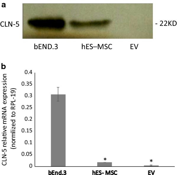
Fig. 4.

CLN-5 protein/gene expression by hES-MSCs and hES-MSC-derived EVs. hES-MSCs were grown in 6-well plates coated with 0.1% gelatin. Cultures of hES-MSCs or bEnd.3 (as a CLN-5 control) were treated with TNF-α (10 ng/ml) for 24 h and extracted for protein or subject to RNA isolation. EVs were prepared from culture supernatants of hES-MCS. (Top) Western blot for CLN-5 protein. (Bottom) qRT-PCR for CLN-5 mRNA. Because total RNA in EVs was too low to accurately detect, qRT-PCR was performed directly in lysed EV extracts following reverse transcription, as described (76). Data are presented as mean ± SE. Each experiment was repeated 3 times. *p < 0.001 compared with bEnd.3
