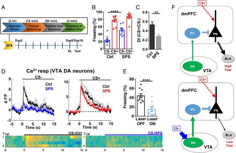
Fig. 7.
Impaired SL and reduced increase in VTA DA signaling in a single prolonged stress model of PTSD. (A) Procedure for establishing the SPS/PTSD model. (B) Freezing levels during CS− [two-way RM ANOVA; interaction, F(1,17) = 4.148, P = 0.0576; stimulus, F(1,17) = 130.1, P < 0.0001; group, F(1,17) = 21.83, P < 0.001; n = 9 mice for control (Ctrl), 10 mice for SPS; CS−, Ctrl vs. SPS, P < 0.0001; CS+, Ctrl vs. SPS, P = 0.15. Bonferroni’s posttest]. (C) Discrimination index in Ctrl and SPS mice (two-tailed unpaired t test, t = 3.470, df = 17; Ctrl vs. SPS, P < 0.01; same set of mice as in B). (D) VTA DA Ca2+ responses during CS− (Left) or CS+ (Right) post-SL in SPS and Ctrl mice [two-tailed unpaired t test; CS−, t = 2.319, df = 10; CS+, t = 0.0158, df = 10; n = 7 mice (Ctrl), 5 mice (SPS); CS−, Ctrl vs. SPS, P < 0.05; CS+, Ctrl vs. SPS, P = 0.99]. Heat maps showed corresponding Ca2+ responses during CS−. (E) Reduced freezing levels during CS− by excitation of VTA DA terminals in the dmPFC after SL in SPS mice (two-tailed paired t test, t = 10.90, df = 9; n = 10 mice; laser off vs. laser on, P < 0.0001). (F) Model of conditioned inhibition. (Left) CS+ (danger signal) leads to high activity in principal neurons (PN) in the dmPFC and high fear via outputs to subcortical regions, such as the basolateral amygdala, (BLA). (Right) After SL, CS− (safety cue) leads to stronger activation of VTA DA neurons, which results in stronger PV neuron activity and reduced PN activity, with low fear expression, in the presence of CS+. The main consequence of SL is enhanced VTA DA neurons from the CS− input, which leads to enhanced transmission from VTA DA neurons to PV neurons and reduced PN neuronal activity (illustrated by thicker lines).

