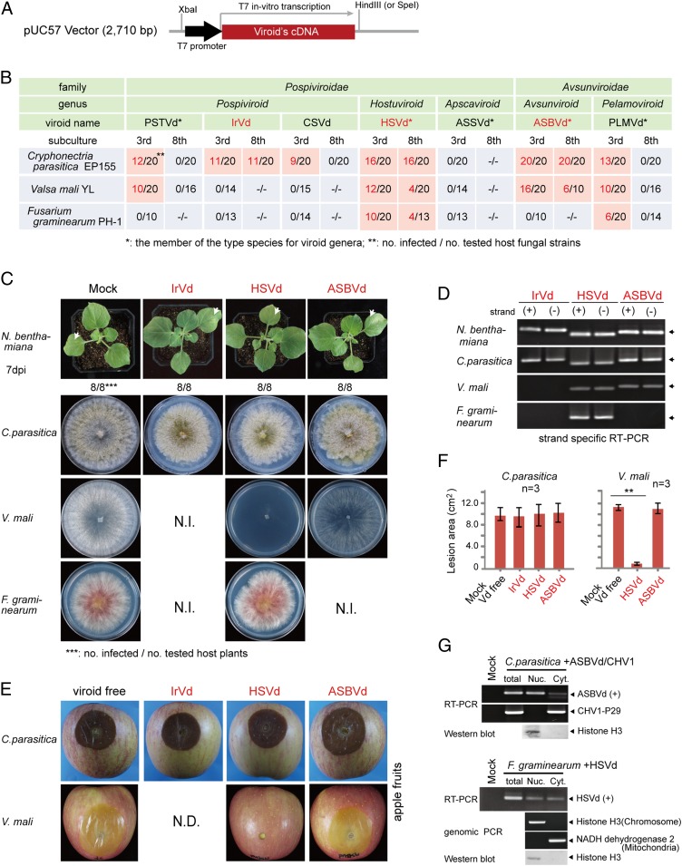
Fig. 1.
Viroid infection and pathogenicity in filamentous fungi. (A) Sequence construct for generating plus (+) strand in vitro transcripts of viroid cDNA clones. (B) Detection of viroid in fungal isolates regenerated from fungal spheroplasts that have been transfected using in vitro transcripts of viroid cDNA clones. Viroid RNA accumulations were detected using RT-PCR at third and eighth fungal subcultures. (C) Phenotypic growth of plants and fungi infected with viroids. Fungi were grown on PDA medium (10-cm plate) for 3 d and photographed. N.I., not infected. (D) RT-PCR analysis of viroid RNA accumulation in the plants and fungi described in C. (E) Fungal virulence assay on apple. Apple fruits were inoculated with mycelial plugs, and fungal lesions were photographed 5 d after inoculation. N.D., no data. (F) Fungal lesion area measured on inoculated apple fruits described in E. **P < 0.01 (Student’s t test). Vd, viroid. (G) Accumulation of viroids in nuclear (Nuc.)- and cytoplasmic (Cyt.)-enriched fractions. Spheroplasts of infected fungi were subjected to cell fractionation using differential centrifugation. Viroid (ASBVd or HSVd) and virus (CHV1) RNAs were detected by RT-PCR, while chromosomal and mitochondrial DNAs (cytosolic DNAs) were detected by genomic PCR. The presence of histone H3 protein in the nuclear fraction was confirmed by Western blotting analysis.

