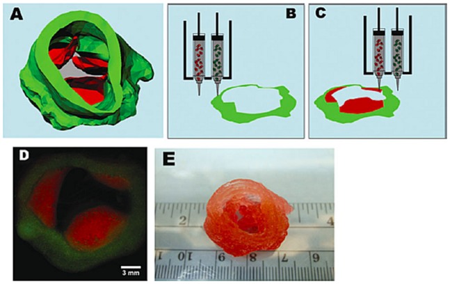
Figure 8.
The 3D bioprinting of an aortic valve conduit. (A) The 3D reconstruction of an aortic valve model. The green color indicates valve root and the red color indicates valve leaflets. Schematic illustration of the 3D bioprinting process using alginate and gelatin as bioinks encapsulated with (B) sinus smooth muscle cells (SMCs) and (C) aortic valve leaflet interstitial cells (VIC) cells. (D) Fluorescent image of two 3D bioprinted layers representing an aortic valve conduit. (E) Macroscopic image of a 3D bioprinted aortic valve conduit. Reprinted from a previous study [196] with permission.

