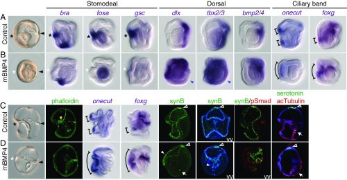
Fig. 2.
Transient overactivation of BMP activity blocks mouth formation and concentrates the larval nervous system ventrally. (A and C) Controls cultured with 125 ng/mL BSA. (B and D) Embryos treated with 125 ng/mL mBMP4. The treatments were performed for 4 h during gastrulation (from 24 to 28 h postfertilization), then washed out. Embryos were fixed at the late gastrula (A and B) or tornaria (C and D) stage for in situ hybridization and immunostaining. BMP activity was monitored using an anti-phospho-Smad1/5/8 (pSmad) antibody. The black asterisks and arrowheads indicate the mouth and the hydropore openings, respectively. The white asterisks denote the disappearance of the mouth and stomodeal gene expression. The blue arrowheads indicate the weaker signal of dlx and tbx2/3 in the most dorsal ectoderm. The yellow arrowhead indicates the pharyngeal muscles in the control tornaria larva. The brackets indicate the expression domains of the onecut and foxg genes in the ventral ectoderm. In mBMP4-treated embryos, synB signals in the postoral ciliary band were concentrated on the ventral side (white arrowheads), while the distributions of the serotonergic neurons (empty arrowheads) and the neurons in the sphincter (arrows) were similar to those of controls. All images are side views (ventral to the left) unless indicated otherwise. VV, ventral view (viewed from the mouth opening side with left and right sides shown). The data represent the phenotypes of most samples (>95%).

