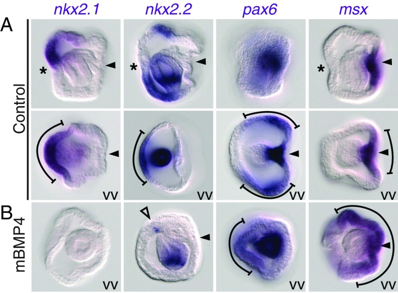
Fig. 4.
Medial-to-lateral organization of the ventral neurogenic ectoderm in P. flava embryos. Shown are expression patterns of nkx2.1, nkx2.2, pax6, and msx in controls (A) and in embryos treated with mBMP4 for 4 h during gastrulation (B). Asterisks and arrowheads indicate the mouth and hydropore openings, respectively. The brackets indicate the ectodermal expression domains. The empty arrowhead indicates the residual ectodermal expression of nkx2.2. Images are either side views or vegetal views (VV). Ventral is to the left in all images.

