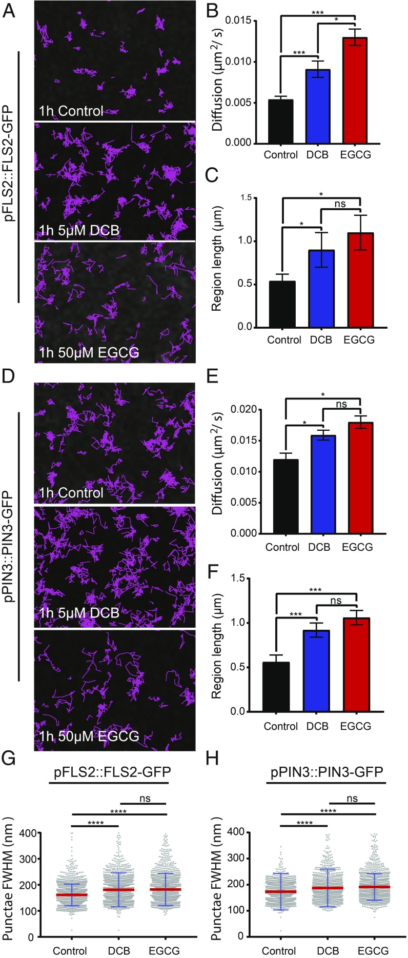
Fig. 5.
Cell wall perturbation alters diffusion rate, constrained region length, and cluster size of FLS2-GFP and PIN3-GFP. DCB was used to perturb cellulose synthesis and EGCG was used to perturb pectin methylation status of hypocotyl epidermal cells. (A–C) Nanodomain characteristics of pFLS2::FLS2-GFP and (D–F) pPIN3::PIN3-GFP in either controls, or after treatment with 5 µM DCB or 50 µM EGCG for 1 h. (A and D) Track length of single particles over 60 s. (B and E) Constrained diffusion rate over 4 s. (C and F) Constrained region length over 4 s. (G and H) FWHM measurement of cluster diameter. Scatter dot plot, red lines show mean value and blue error bars show SD. There was a significant increase or trend toward increase in all nanodomain characteristics for both proteins after cell wall perturbration. *P < 0.05, ***P < 0.01, ****P < 0.001; ns, not significant.

