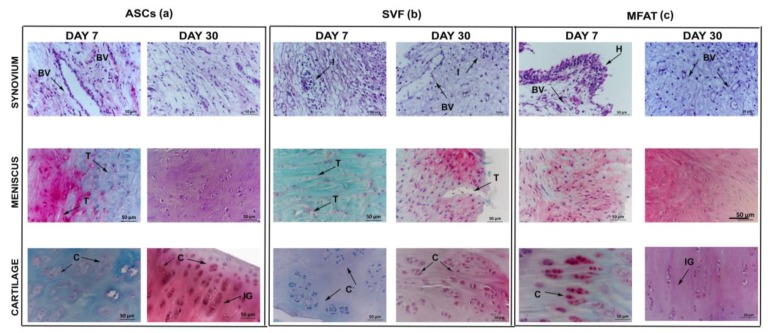
Figure 5.
Representative micrographs of histological assessment of ASCs (a), SVF (b), and MFAT (c) in the synovial membrane; the meniscus and the cartilage were stained with Hematoxylin/Eosin and Safranin-O/Fast Green, respectively, at 7 and 30 days. Scale bar: 50 µm. Black arrows: indications of some histological details in tissue specimens. BV: blood vessels; I: inflammatory processes; H: hyperplastic and hypertrophic processes in the synovial membrane; T: tear presence in meniscus C: cell clones within the extracellular matrix in cartilage; F: fibrillation processes; IG: isogenic groups.

