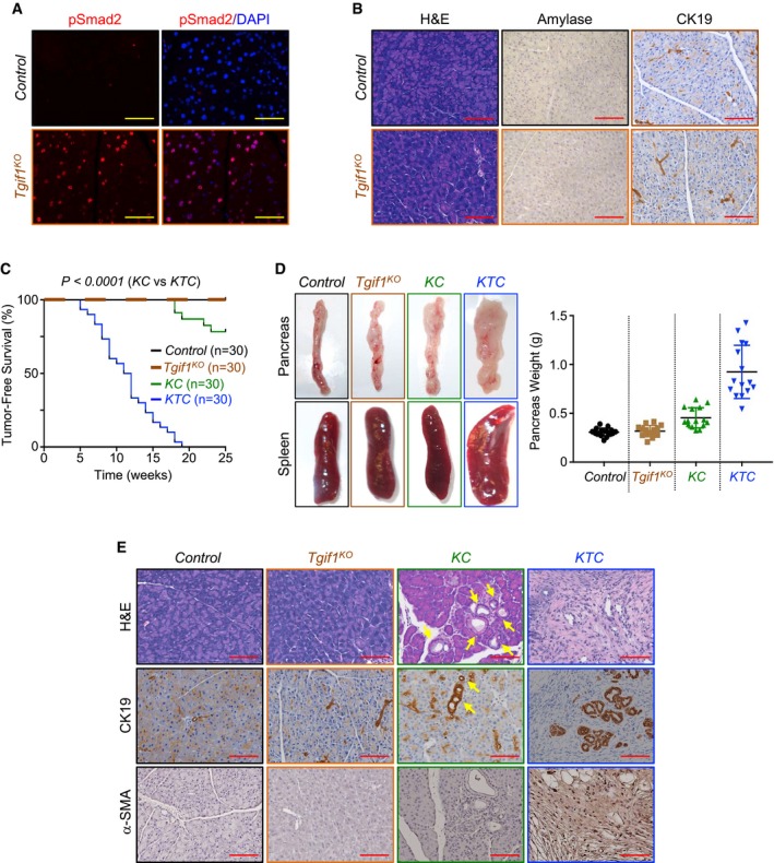
Formalin‐fixed paraffin‐embedded (FFPE) sections from control or Tgif1
KO mice were immunostained with anti‐pSmad2 antibody and revealed by immunofluorescence (IF) and DAPI. Representative pictures at 40× are shown (n = 30). Scale bars, 100 μM.
FFPE pancreatic sections from control or Tgif1
KO mice were stained with hematoxylin and eosin (H&E) or immunostained with antibodies to CK19 or Amylase and revealed by IHC. Representative pictures at 20× are shown (n = 30). Scale bars, 200 μM.
Kaplan–Meier survival analysis of control, Tgif1
KO, KC, and KTC mice. A regular mosaic two‐color line was used to discriminate between control and Tgif1
KO mice.
Pictures of whole pancreas and spleen tissues from control, Tgif1
KO, KC, or KTC mice (left). Weight of pancreas was measured and presented as dot plot (n = 30).
FFPE pancreatic sections from control, Tgif1
KO, KC, or KTC were stained with H&E or subjected to IHC analysis using antibodies to CK19 or α‐SMA. Yellow arrows indicate PanINs in KC mice. Representative pictures at 20× are shown (n = 30). Scale bars, 200 μM.

