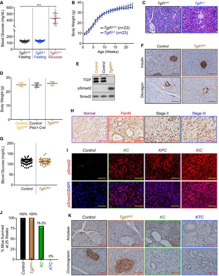
Tgif1
+/+ or Tgif1
−/− mice were fasted for 6 h and blood glucose levels were measured using the ReliON method. Mice injected with glucose at 2 g/kg body weight were used as controls. ***P < 0.001; based on a two‐tailed Student's test.
Body weight of Tgif1
+/+ or Tgif1
−/− mice was recorded every week for 25 weeks.
FFPE pancreatic sections from Tgif1
+/+ or Tgif1
−/− mice were stained with H&E. Representative pictures at 20× are shown (n = 23). Scale bars, 200 μM.
Body weight of control or Tgif1
KO mice was measured at the age of 25 weeks.
Total lysates from pancreas of control or Tgif1
KO mice were pooled and analyzed by immunoblotting using antibodies to TGIF1, pSmad2, and Smad2 used as control (n = 6).
FFPE pancreatic sections from control or Tgif1
KO mice were immunostained with antibodies to Insulin or Glucagon and revealed by IHC. Representative pictures at 20× are shown (n = 30). Scale bars, 200 μM.
Fasting blood glucose of control or Tgif1
KO mice was measured as described in (A).
Expression of TGF‐β1 in tissue microarrays of human PDAC samples was analyzed by IHC. Representative pictures at 40× are shown. Scale bars, 100 μM.
FFPE pancreatic sections from control, KC, KPC or KIC mice were immunostained with anti‐pSmad2 antibody and revealed by IF and DAPI. Representative pictures at 40× are shown (n = 7–30). Scale bars, 100 μM.
Tumor‐free survival of control, Tgif1
KO, KC and KTC mice at 25 weeks of age, time at which 100% of KTC mice succumbed to PDAC.
FFPE pancreatic sections from control, Tgif1
KO, KC or KTC mice were immunostained with antibodies to chromogranin A or Amylase and revealed by IHC. Representative pictures at 40× are shown (n = 30). Scale bars, 100 μM.

