-
A
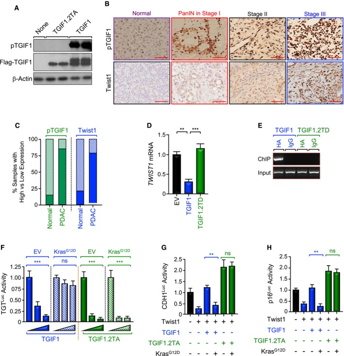
Specificity of anti‐phospho‐TGIF1 (pTGIF1) antibody. Cell extracts from Tgif1
−/− MEFs cells reconstituted with empty vector (EV), wild‐type (TGIF1) or non‐phosphorylated form (TGIF1.2TA) of Flag‐TGIF1 were immunoblotted with anti‐phospho‐TGIF1 or β‐Actin as a loading control.
-
B, C
Expression of pTGIF1 and Twist1 in human tissue microarrays of human PDAC samples was analyzed by immunohistochemistry. Representative pictures at different stages (40×) are shown. The percentages of samples with high versus low expression of pTGIF1 and Twist1 in normal tissues versus PDAC tissues are shown. Scale bars, 100 μM.
-
D, E
MIAPaCa‐2 cells were transfected with TGIF1 or TGIF1.2TD expression vectors and selected with G418, and resistance colonies were pooled (n = 6). Expression of Twist1 was examined by qRT–PCR (D). Binding of TGIF1 to the Twist1 promoter was examined by ChIP and agarose gel.
-
F
BxPC3 cells were transfected with TGTLuc together with TGIF1 or TGIF1.2TA in the presence or absence of KrasG12D. Luciferase activity was measured 48 h following transfection and normalized on the basis of co‐transfected Renilla luciferase (n = 6).
-
G, H
BxPC3 cells were transfected with CDH1Luc or p16Luc reporter together with TGIF1 or TGIF1.2TA in the presence or absence of KrasG12D, and luciferase activity was measured 48 h following transfection as described in (E) (n = 6).
Data information: For (D, F, G, and H), data are expressed as mean ± SEM. **
P < 0.01; ***
P < 0.001; ns: not significant; based on a two‐tailed Student's test.

