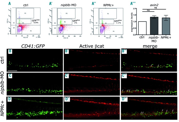
Figure 4.
The canonical Wnt pathway is hyper-activated specifically in hematopoietic stem cells. (A-A”) Fluorescence-activated cell sorting analysis of CD41:GFPlow cells from controls (A), nipblb-MO (A’) and NPMc+ mRNA (A”) injected embryos at 3 days post-fertilization (dpf) and quantitative reverse transcriptase polymerase chain reaction analysis of axin2 expression on sorted cells (A”’). (B-D”) Immunofluorescence staining with green fluorescent protein (GFP) for hematopoietic stem cells. (B-D) and Active β-catenin for Wnt activation (Active β-cat) (B’-D’) antibodies. Merging of the two signals (B”-D”) showed an increased number of GFP/Active β-cat double-positive cells (arrows) at 3 dpf in the caudal hematopoietic tissue of embryos injected with nipblb-MO or NPMc+ mRNA in comparison to the number in controls. Images were processed as described in the Online Supplementary Methods. The scale bar represents 100 μm. *P<0.05. ns: non-significant; ctrl: control; MO: morpholino.

