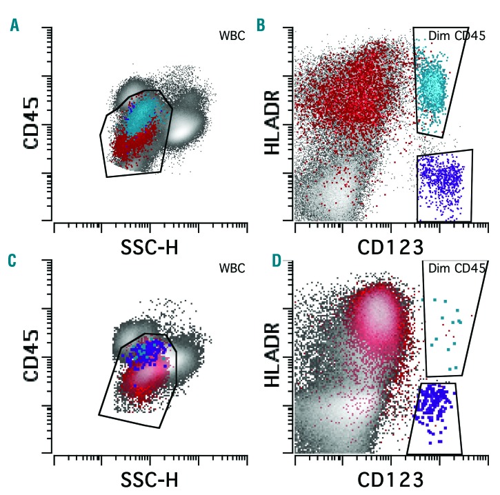
Figure 1.
Loss of plasmacytoid dendritic cell (PDC) differentiation in acute myeloid leukemia (AML). PDC from a control subject (A and B) reside in the CD45 dim/low side scatter gate between blasts and monocytes, overlapping with basophils (A). PDC express high levels of CD123 and HLA-DR and can be easily separated from blasts and basophils (B). In AML with morphological disease (≥20% blasts) (C and D), PDC are markedly reduced. Red: CD34 positive blasts; blue: PDC; purple: basophils.

