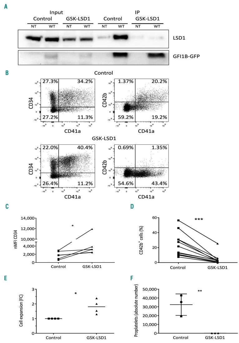
Figure 3.
Chemical disruption of the GFI1B-LSD1 interaction by GSK-LSD1 recapitulates GFI1BQ287* hallmarks in vitro. (A) Western blot on co-immunoprecipitated (IP) LSD1 after GFP-trap bead pulldown in dimethylsulfoxide (DMSO) control or GSK-LSD1-treated MEG-01 cells transduced with GFI1B-GFP (WT). Non-transduced (NT) cells were used as a negative control. The upper panel shows LSD1 (~90 kDa) and the lower panel GFI1B variants-GFP (~58 kDa). The left side of the blot shows LSD1 and GFI1B-GFP expression in the input samples. (B) Cell surface expression of megakaryocyte-associated markers CD34, CD41a, and CD42b, to measure megakaryocyte maturation. The presented results are from megakaryocytic cultures 2 days after addition of DMSO control or 4 μM GSK-LSD1. (C) CD34 normalized median fluorescence intensity (nMFI) from CD34+/CD41a+ megakaryoblasts. (D) Percentage of CD42b positivity on CD41a+ megakaryoblasts. The connecting lines (C-D) indicate which samples are from the same experiment. (E) Expansion of CD34+/CD41a+ megakaryoblasts during 2 days of DMSO control or GSK-LSD1 treatment. (F) Absolute number of proplatelet-forming megakaryocytic cells per view (based on Online Supplementary Figure S4A,B) 6 days after addition of DMSO or 4 μM GSK-LSD1 (n=6). *P<0.05, ***P<0.001.

