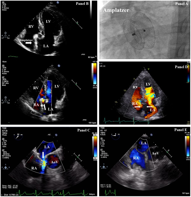
Figure 1.
(A) Fluoroscopy (antero-posterior projection) exhibiting correct placement of 34-mm Amplatzer Septal Occluder device. (B) Transthoracic echocardiography (four-chamber projection) showing malalignment of the septal occluder device (arrow) with the interatrial septum. (C) Transoesophageal echocardiography demonstrating left to right interatrial shunt. (D) Transoesophageal echocardiography after removal of the atrial septal defect septal occluder device and patch closure of the secundum atrial septal defect. AO, aorta; LA, left atrium; LV, left ventricle; RA, right atrium; RV, right ventricle.

