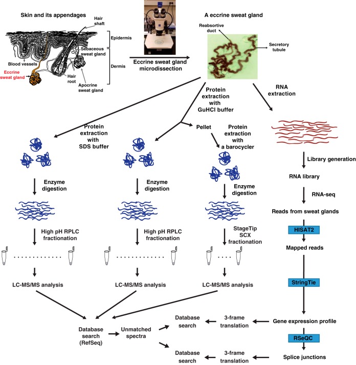
Fig. 1.
Schematic diagram for the research strategy. Human eccrine sweat glands were microdissected from human skin. Top, the left side of the figure shows a diagrammatic representation of skin and its appendages. Top, the right side of the figure shows a micrograph of an eccrine sweat gland dissected from the skin using stereomicroscope. Transcriptome and proteome analyses were conducted after the extraction of proteins and RNAs, respectively. For transcriptome analysis, the extracted mRNAs were used for the library preparation and subjected to the RNA-seq. The RNA-seq data were analyzed using HISAT2, StringTie and RSeQC. For proteome analysis, the extracted proteins were digested with Lys-C followed by trypsin. The resulting peptides were pre-fractionated by either bRPLC or an SCX StageTip followed by LC-MS/MS analyses. Acquired mass spectrometry data were subject to a reference protein database and the resulting unmatched spectra were subject to another round of a search against customized databases generated by 3-frame translation of either the RNA-seq or splice junction data.

