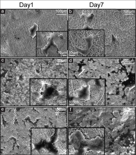
Figure 3.

Scanning electron microscopy images of chondrocyte cells seeded on electrospun (a and b) polyhydroxybutyrate, (c and d) polyhydroxybutyrate-chitosan, and (e and f) polyhydroxybutyrate-chitosan/3% Al2O3 nanowires, left column at day 1 and right column on day 7
