Figure 1.

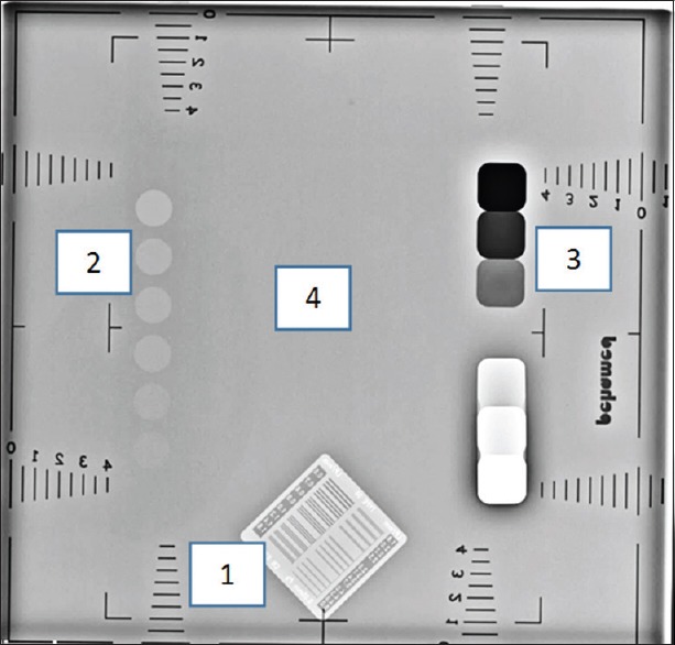
The radiographic image of DIGRAD A + K digital radiography phantom. Location of the different parts of the phantom: (1) spatial resolution, (2) low contrast detectability, (3) contrast dynamic range and (4) noise are seen

The radiographic image of DIGRAD A + K digital radiography phantom. Location of the different parts of the phantom: (1) spatial resolution, (2) low contrast detectability, (3) contrast dynamic range and (4) noise are seen