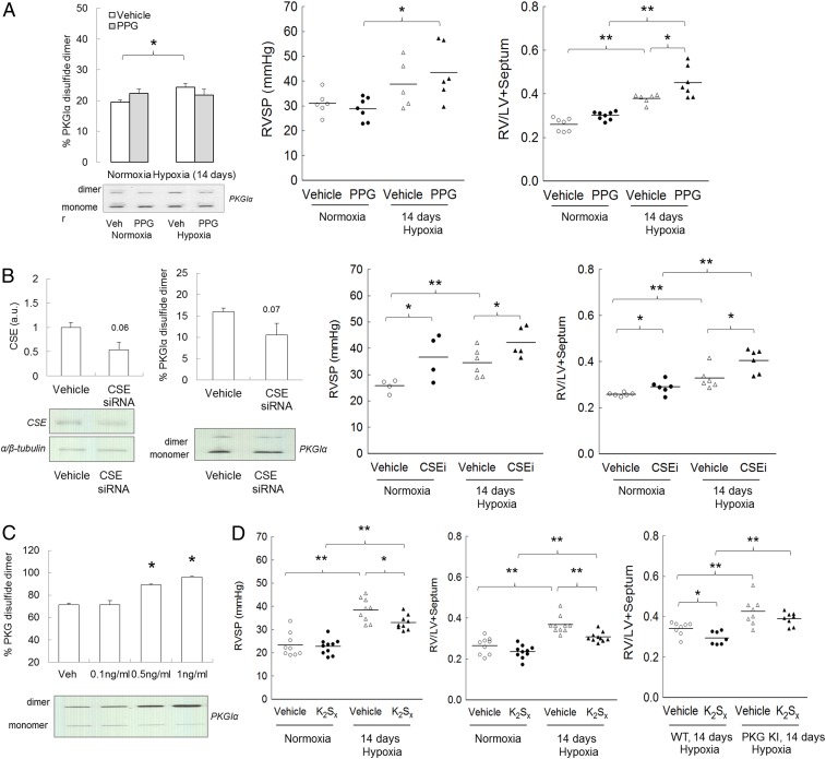
Fig. 3.
Effect of CSE inhibition and polysulfides in hypoxia-induced PH. (A) Pulmonary disulfide PKGIα expression, RV pressure, and RV to left ventricle + septum (LV+S) ratio in C57BL/6 mice subjected to either normoxia or chronic hypoxia for 14 d with or without CSE inhibitor l-PPG (50 mg/kg/d). (B) CSE protein expression and disulfide PKGIα level in lungs of mice treated with CSE siRNA (1.3 mg⋅kg−1⋅d−1); RV pressure and RV to LV+S ratio in WT mice subjected to either normoxia or chronic hypoxia for 14 d with or without CSE siRNA (1.3 mg⋅kg−1⋅d−1). (C) Disulfide PKGIα formation in response to persulfides donor K2Sx treatment in human pulmonary artery smooth muscle cells. (D) RV pressure and RV to LV+septum ratio in C57BL/6 mice subjected to either normoxia or chronic hypoxia for 14 d with or without persulfides donor K2Sx (2 mg⋅kg−1⋅d−1); RV to LV+S ratio in WT or KI mice subjected to chronic hypoxia for 14 d with or without K2Sx (2 mg⋅kg−1⋅d−1). *P < 0.05, **P < 0.01 versus control; n = 5 to 7 per group. In some cases, the aspect ratio of the original immunoblots was altered to enable a concise multipanel figure with a consistent presentation style; the original uncropped representative images of these immunoblots are also available in SI Appendix, Fig. S10.

