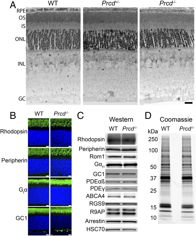
Fig. 2.
Prcd−/− mice develop all retinal layers and have normal localization and abundance of outer segment proteins. (A) Retinal morphology of WT, Prcd+/−, and Prcd−/− mice at P21. The 500-nm retinal cross-sections embedded in plastic were stained by Toluidine blue and analyzed by light microscopy (Scale bar, 20 µm). (B) Immunostaining of retinal cross-sections from WT and Prcd−/− mice at P21 with antibodies against representative ROS proteins indicated in the panel (green). Nuclei are stained with Hoescht (blue) (Scale bars, 10 µm). (C) Western blotting of representative outer segment proteins from retinal lysates of WT and Prcd−/− mice at P21. Samples are normalized by total protein. (D) A Coomassie-stained SDS/PAGE gel loaded with equal amounts of outer segments purified from WT and Prcd−/− mice at P21. Outer segments were purified in the dark using a density gradient, and samples were normalized by their content of rhodopsin. Data for all panels are taken from one of at least three independent experiments.

