Fig. 3.
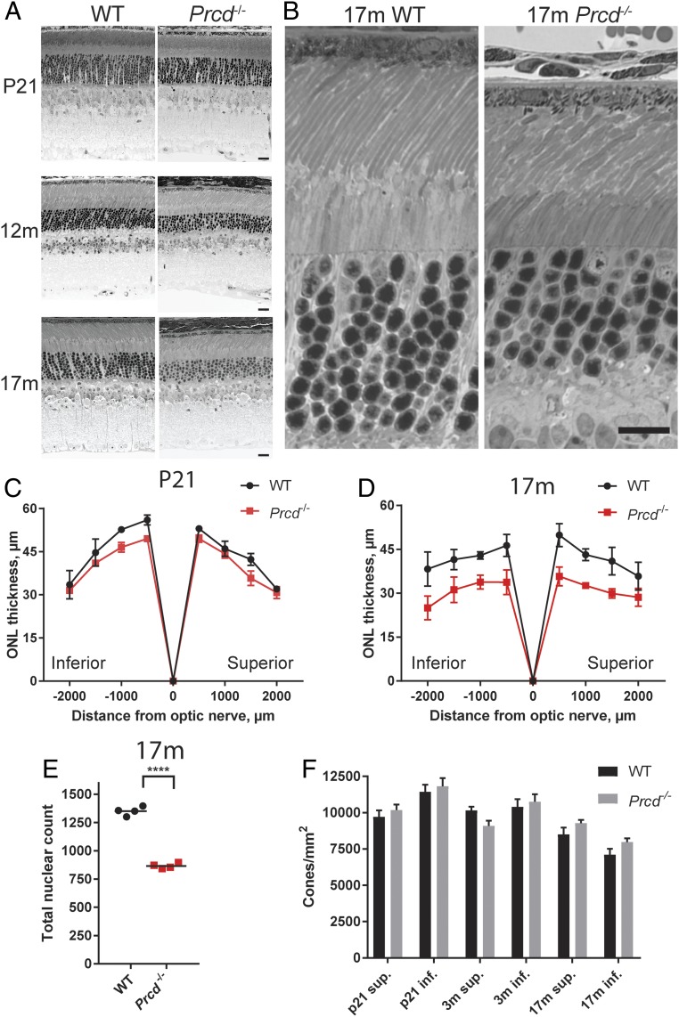
Rod photoreceptors of Prcd−/− mice undergo slow degeneration, while cones survive. (A) Representative images of retinal cross-sections from WT and Prcd−/− mice taken at P21, 12 mo, and 17 mo showing the progressive loss of the ONL in Prcd−/− mice (Scale bars, 20 µm). (B) Representative, high-magnification images of WT and Prcd−/− mouse outer retinal layers at 17 mo of age (Scale bar, 20 µm). (C and D) Spider diagrams representing the thickness of the ONL in the inferior and superior retinas of WT and Prcd−/− mice at various distances from the optic nerve head. Measurements were conducted with P21 and 17-mo-old mice; one retina from each of four mice was analyzed for each genotype. Data are shown as mean ± SD determined for each location. (E) The reduction of the number of photoreceptor nuclei in 17-mo-old Prcd−/− mice. Nuclei were counted and summed from eight 100-µm-wide segments centered at each of the eight locations analyzed in the spider diagrams in D. Data are shown as mean ± SD; ****P < 0.0001, two-tailed t test. (F) Cone photoreceptor density in retinal flat mounts from WT and Prcd−/− mice of indicated ages. Cell count was performed after staining cones with peanut agglutinin (PNA), as shown in SI Appendix, Fig. S3. No significant difference (P > 0.05) was found between WT and Prcd−/− mice at any age. A two-way ANOVA with Bonferroni correction for multiple comparisons was used for statistical analysis.

